Atrial Fibrillation
We can help.
Atrial Fibrillation is when the heartbeat in the filling chambers of the heart (atria) goes out of rhythm. Atrial Fibrillation causes the pumping of the heart to be inefficient and increases the risk of stroke. Your heartbeat may feel fast and irregular, you may feel tired or short of breath, or you may not notice it.
Causes of Atrial Fibrillation:
These include: high blood pressure, weak or stiff heart, heart valve problems, poor lifestyle choices, or problems with wiring of the heart.
Risks due to atrial fibrillation:
- Stroke: there is a risk of a blood clot going from the heart to the brain causing a stroke. This risk increases depending on the number of risk factors for stroke. Theses risk factors include: female gender, weak or stiff heart, hypertension, diabetes, previous strokes, age greater than 65 years.
- Heart Failure: Atrial fibrillation can also cause fluid to build up behind the heart (heart failure).
Investigations:
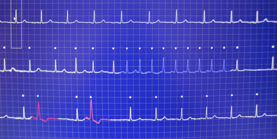
Resting Electrocardiograph (ECG)
A Resting ECG at Cardiology Specialists is an electrical tracing of your heart. Electrodes are placed on the skin of your arms, legs and across chest to measure electrical activity of the heart. It gives information about heart rate, evidence of old heart attacks, thickening of heart muscle, or heartbeat problems such as extra (ectopic) beats or atrial fibrillation.
24 hour Ambulatory Holter Monitor (Heart rate monitor)
 24 hour Holter Monitor at Cardiology Specialists is where a continuous heart rate tracing is obtained to measure relationship between symptoms and heartbeat. Electrodes are applied to your chest under your clothes by trained staff to measure continuous measurement of heart rate and rhythm over 24 hours. The electrodes are attached to a small portable recorder. You can mark the recording at any time you have heart flutters or other symptoms. It is important to keep a diary so your symptoms can be compared to your heart rhythm at same time. The 24 hour Holter Monitor at Cardiology Specialists is useful to detect different types of palpitations including: extra heart beats (ectopic beats), fast regular heart beats (supraventricular tachycardia (SVT), irregular heart rhythm (atrial fibrillation), and ventricular tachycardia (VT). Patients with intermittent atrial fibrillation are at similar risk for stroke compared to patients in atrial fibrillation all the time.
Echocardiography
Echo at Cardiology Specialists uses ultrasound to measure function of heart chambers and heart valves. Echo is also called Cardiac Ultrasound, which uses high frequency sound waves and is similar to a gall bladder or pregnancy ultrasound but is instead focused on the heart. Professor Hamid Ikram pioneered the introduction of Echo to Canterbury. At Cardiology Specialists, Echo is performed by a specially trained technician who moves a plastic transducer on the skin of the chest wall to obtain pictures of the heart chambers and valves. Echo (cardiac ultrasound) is useful for diagnosing weakened heart muscle, old heart attacks, heart valve narrowing or leaking, thickening of heart muscle, holes between heart chambers, or fluid in the sack around the heart.
Treatments for Atrial Fibrillation
Electrical Cardioversion
Dr Dougal McClean performs Electrical Cardioversion for Atrial Fibrillation. Under a General Anaesthetic, a synchronised electric shock is performed to the outside of the chest to reset the heart. Patients need to take blood thinners for at least 3 weeks prior to Cardioversion, and for 4 weeks afterwards, to prevent the risk of stroke.
Dr Dougal McClean is an affiliated provider with Southern Cross for Electrical Cardioversion under a General Anaesthesia.
Lifestyle changes such as losing weight, decreasing alcohol, and improving blood pressure control are important.
Patients will receive medications to prevent further episodes of fibrillation and lessen risk of stroke. It is important that these medications don’t cause side effects.